

Fluorescence spectra, like their enhances, absorption and excitation spectra, are usually very broad – the sign happens over many wavelengths. In consequence, when quite a few probes are getting used concurrently, sign from one probe might be detected within the spectral area of one other probe. This drawback is termed “crosstalk” (or “spillover” in move cytometry). Determine 1 illustrates a number of fluorescent labels being excited with a single laser. It could be logical to cut back crosstalk by selecting fluorophores which are extra spectrally separated, however this isn’t at all times an possibility, particularly when utilizing fluorescent proteins.
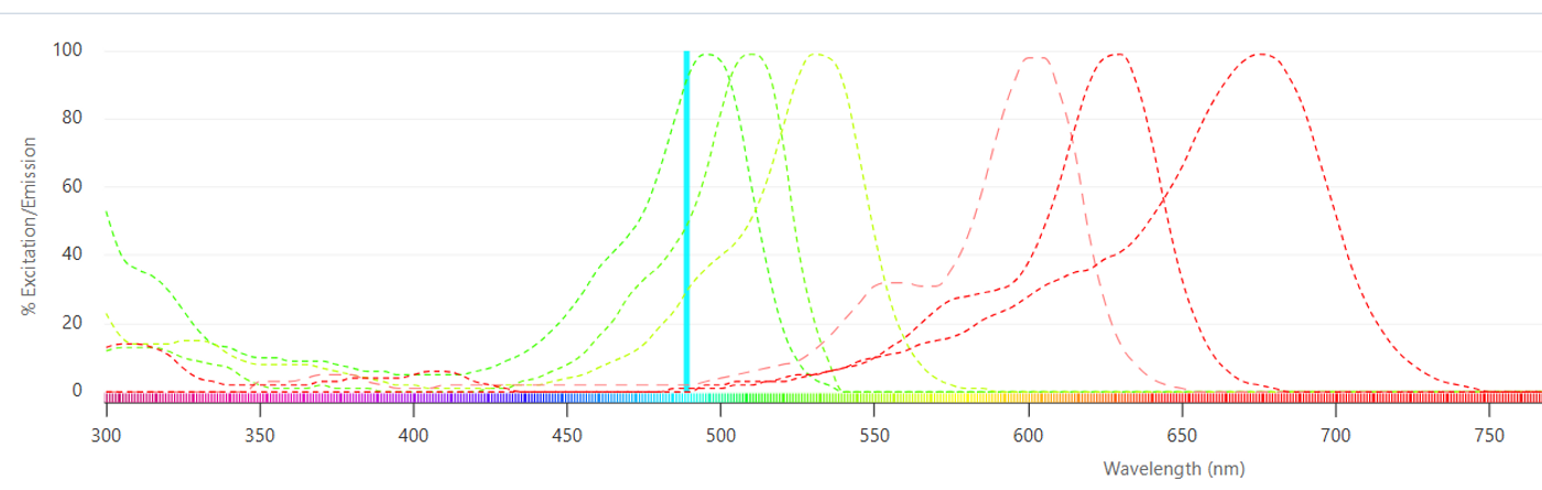
Determine 1. Quite a few fluorescent probes might be excited with a single laser (blue line).
In confocal microscopy, laser excitation gentle is raster scanned throughout the pattern to induce the fluorescence sign. The fluorescence is collected by the target lens and descanned by way of a pinhole to cut back sign from above and beneath the aircraft of curiosity. Commonplace confocal microscopes then direct the fluorescence sign by way of a sequence of optical filters (dichroic beamsplitters and band-pass filters) into single-element detectors (typically PMTs) to learn off the sign pixel-by-pixel. As seen in Determine 2, even with optimized band-pass filters, there may be important crosstalk between fluorescence alerts. Utilizing bandpass filters additionally significantly reduces the sign depth of every fluorophore as a result of out-of-band gentle is just not detected.
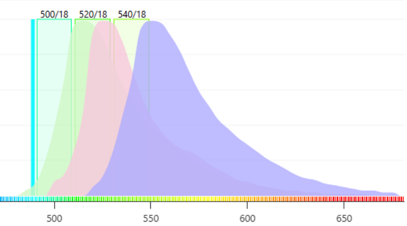
Determine 2. It’s tough to acquire “pure” alerts from every fluorophore utilizing optical bandpass filters.
To unravel this drawback, the primary spectral confocal microscopes had been launched within the mid-2000s, beginning with the Zeiss Meta and rapidly adopted by choices from different main microscope firms. These programs have been improved over time and use quite a lot of strategies to create the spectra. Whatever the technique, all the devices can produce a full emission spectrum at every pixel. This spectrum is a sum of the emission from all the contributing fluorophores in that pixel (Determine 3).
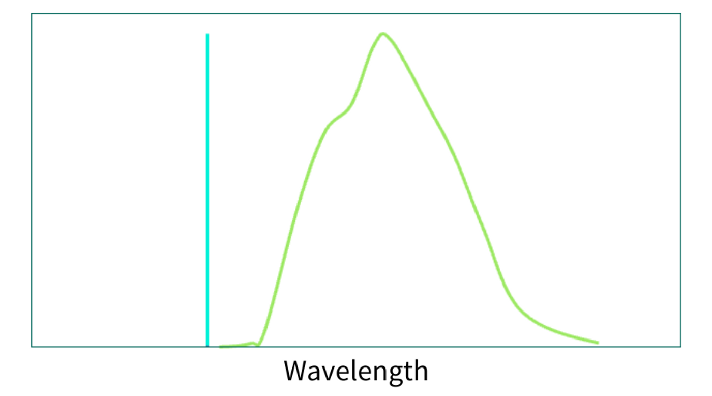
Determine 3. Instance of a mixed spectrum of the three spectra above.
Linear Spectral Unmixing
Knowledge processing consists of linear spectral unmixing to find out the proportion of every fluorophore’s contribution to the spectrum. Reference spectra are required for the unmixing. These might be acquired individually (or in multi-labeled subsets) as management pictures, or in spectral libraries of frequent fluorophores offered by microscope producers. Given the recognized spectra and the measured spectrum, one can calculate the contributions of every (Determine 4).
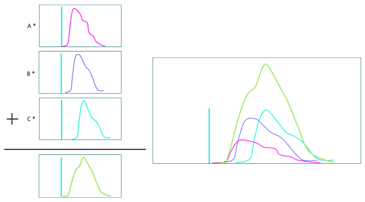
Determine 4. Left: Contributions of every spectrum (A,B,C) are calculated. Proper: The three spectra with the right coefficients mix to kind the measured spectrum.
One benefit to Linear Spectral Unmixing is that it may be used to eradicate autofluorescence sign from the picture. Autofluorescence spectra rely upon the tissue sort and may come from quite a lot of intrinsic fluorophores together with NADH, FAD, chlorophyll, lipofuschin, retinals, melanin, and many others. and may come up as a fixation artifact. Generally an autofluorescence spectrum of an unlabeled pattern is acquired as one of many reference spectra. This contribution can then be hidden from the labeled picture.
Linear Spectral Unmixing requires that there be no less than one detection channel per fluorophore and that the fluorophore spectra usually are not similar.
The {Hardware}
There are lots of strategies to resolve the spectral options of the fluorescence sign (determine 5). The unique technique, nonetheless extensively utilized in microscopy and move cytometry, includes a sequence of dichroic and bandpass filters and particular person detectors for every wavelength band as described above. On condition that the transmission (or reflectance) of a filter is often about 95%, the sign power degrades because it goes by way of the chain of dichroics and detectors. This limits the sensible variety of detectors to about 6. This limitation prompted microscope firms to give you progressive spectral design options beginning within the early-to-mid 2000s.
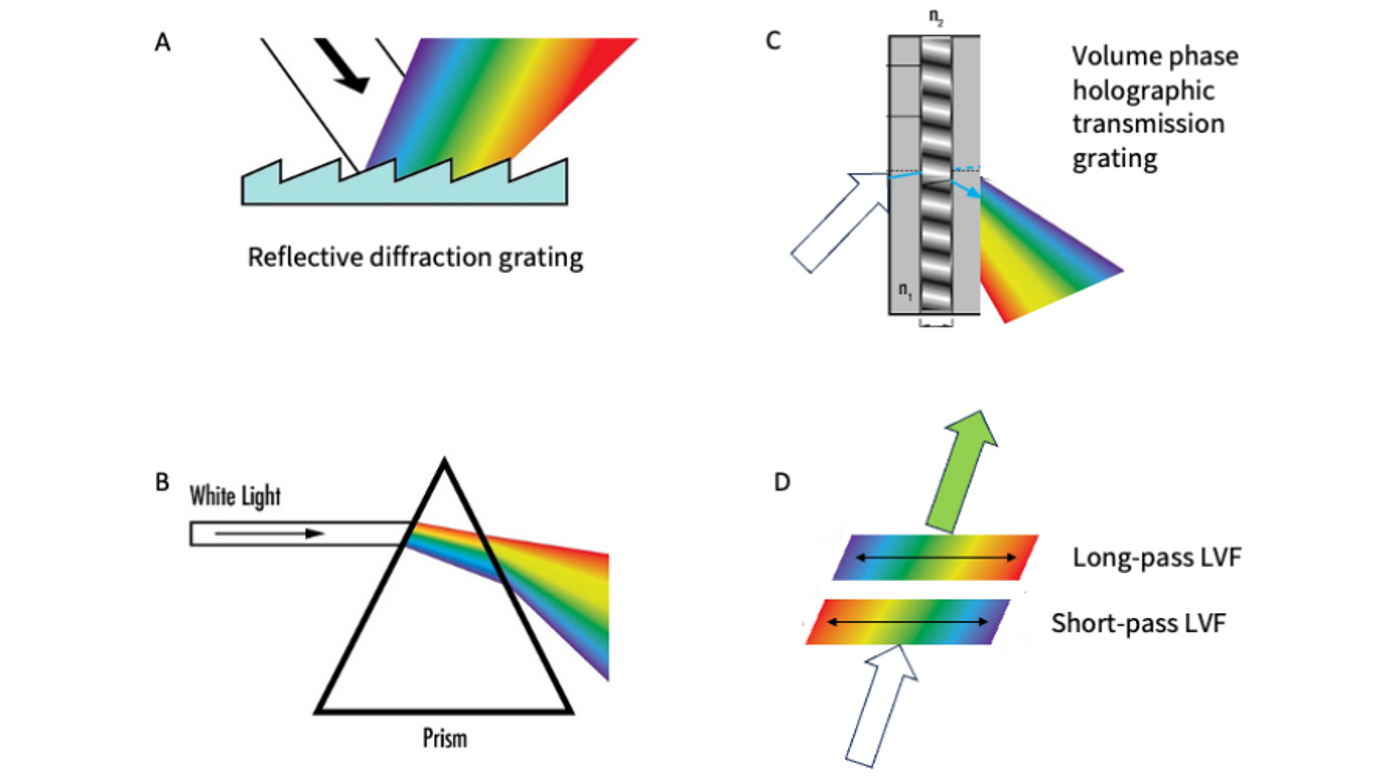
Determine 5. Totally different strategies of manufacturing spectral dispersion utilized in trendy microscopes.
The preliminary method by Zeiss within the 510 Meta instrument within the early 2000s was to make use of a reflective diffraction grating and a linear detector array. Diffraction gratings use the interplay gentle with microscopic grooves on the floor to induce spectral separation (Determine 5a). The wavelength array (rainbow) is targeted onto a linear array of photomultiplier tubes, enabling the simultaneous acquisition of all wavelength bands. This method has been refined over time, including extra optics for beam recycling, growing the sensitivity and variety of channels within the array (to 32) and by splitting off the low (near-UV) and excessive (NIR) wavelength edges of the spectrum to single-element detectors permitting them to be run at increased achieve settings to account for sensitivity drop-off at these wavelengths. This “Quasar” detector meeting is the center of the LSM980 and gives a spectrum from 380-720 nm with 34 complete channels, with 10 nm spectral decision. This design permits a quick, single-shot spectrum at every pixel. All channels are collected concurrently with out scanning or transferring elements. This instrument has been used to picture 10 fluorophores (9 fluorescent proteins and a calcium indicator) simultaneously in living mice.
Leica employs a prism to attain spectral dispersion of the emission sign (Determine 5b). Prisms very effectively separate colours by refraction. Through the years, Leica has refined the prism design and detector configurations. The Stellaris detector module, used of their newest confocal microscopes, makes use of 5 excessive effectivity detectors that allow scanning within the 410-850 nm vary with adjustable decision. Leica not too long ago printed an illustration of unmixing a 15-plex fluorophore-labelled mouse tumor tissue sample.
Olympus (now Evident) makes use of a mix of dichroic filters and quantity section holographic transmission gratings (determine 5c) of their FluoView 3000 Lightpath confocal. Holographic gratings are manufactured from a movie of fabric with alternating refractive indexes (perpendicular to the floor) sandwiched between glass home windows. The refractive index modulation within the movie causes diffraction of the incoming beam into its spectral parts. Their system design makes use of dichroic beamsplitters to separate the spectral alerts into varied channels so completely different spectral areas might be scanned concurrently with completely different resolutions and achieve settings.
Nikon makes use of pairs of linear variable filters to allow customized bandpass configurations or wavelength scanning (determine 5d) in its DUX-VB detector module. Linear variable filters are optical filters the place the spectral response modifications linearly alongside the size of the filter. Utilizing a mix of long-pass and short-pass filters, band-passes with customized widths and edge positions might be created. By concurrently transferring the short- and long-pass filters throughout the beam, wavelength scanning might be achieved within the 400-750 nm vary, whereas concurrently altering the spectral decision if desired.
Supporting Your Analysis
As mentioned, there are lots of intelligent approaches to reaching spectral separation, flexibility in decision, pace and sensitivity in these programs. When mixed with different strategies like cyclical immunofluorescence techniques or fluorescence lifetime imaging, high-level multiplexing of scores of probes is turning into commonplace, unlocking a wealth of recent info for the trendy biologist.
FluoroFinder’s Microscopy Spectra Viewer and Panel Builder will help you visualize and choose optimum fluorophore mixtures and minimizing spectral crosstalk, making certain cleaner and extra dependable knowledge. By streamlining the method of designing complicated panels and providing complete spectral knowledge, FluoroFinder empowers scientists to maximise the potential of recent imaging programs and deal with the challenges of high-level multiplexing with confidence.
Trending Merchandise